How Indian scientists are mapping the brain’s last frontier
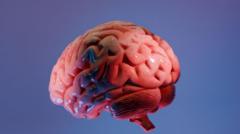
Scientists at the Sudha Gopalakrishnan Brain Centre at the Indian Institute of Technology, Madras, say they have created the world's most detailed three-dimensional atlas of the human brainstem at cellular resolution. Called Anchor (Atlas of Neurochemical Characterisation of the Human Brainstem with 3D Reconstruction), the digital map allows scientists to move seamlessly from whole-brain MRI scans down to individual nerve cells, addressing a long-standing gap between medical imaging and cellular pathology. The achievement matters because the brainstem controls vital functions such as breathing, heartbeat, sleep and movement, yet its densely packed architecture has frustrated efforts to map it in detail.
The atlas combines more than 500 tissue sections from foetal, childhood and adult brains, and was built using high-resolution microscope images rather than costlier molecular techniques. It identifies more than 200 clusters of brain cells and nerve pathways, using eight chemical markers to distinguish different cell types. Experts have praised the work: neuroscientist Shubha Tole called it a "visionary programme" that puts India at the international table, while Harvard-affiliated neuropathologist Rebecca Folkerth, who collaborated on the project, said it realises what she had "dreamed of" early in her career — matching brain scans to microscopic anatomy. Pathologists currently examine only a tiny fraction of the brain's 86 billion neurons, typically 15 to 20 sections when diagnosing conditions such as Alzheimer's disease.
In short
- Indian scientists have built the most detailed 3D brainstem atlas yet.
- It links whole-brain MRI scans to individual nerve cells.
- Built from 500-plus tissue sections, identifying over 200 cell clusters.
Explained simply
The brainstem is the lower part of the brain that controls essential bodily functions such as breathing, heartbeat and sleep. Understanding its structure in detail has long been challenging because it is densely packed and complex, even though this knowledge could help diagnose serious diseases including Alzheimer's.
Researchers at the Indian Institute of Technology in Madras have created a detailed three-dimensional digital map of the human brainstem by studying tissue from foetuses, children and adults. This atlas allows doctors and scientists to match what they see in medical scans with the actual microscopic structure of brain cells, bridging a gap that has existed in medical science for years.
Currently, doctors examining brains for disease typically look at only a small number of tissue samples—around 15 to 20 sections from the brain's roughly 86 billion nerve cells. A better understanding of brainstem structure could improve how doctors diagnose and treat conditions affecting this vital region of the brain.