World news and discussion on TalkFuse.
College students discover new muscle attachment that could improve hip surgery
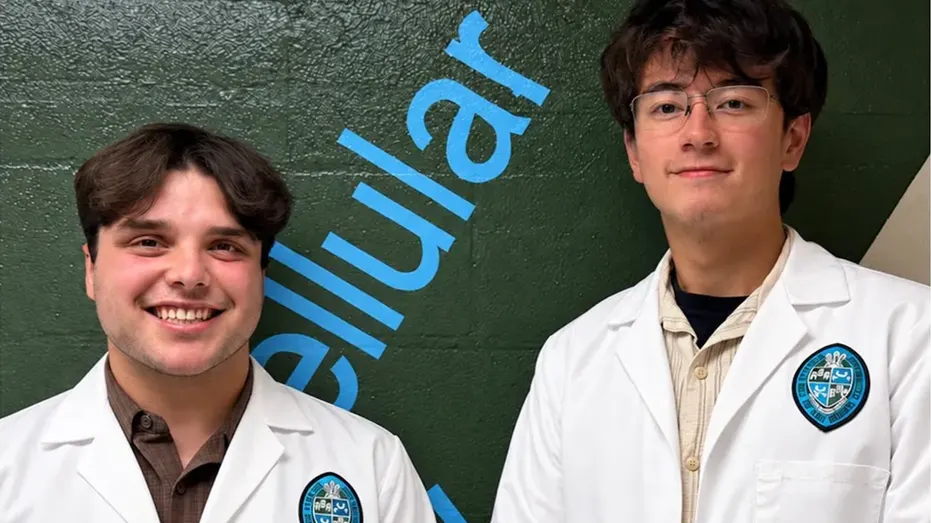
Two Tulane University undergraduates, Dominic Roy and Cooper Bruce, identified a previously undescribed muscle attachment in the front of the hip while working in a cadaver lab. Their professor, anatomy specialist R. Shane Tubbs, could not initially identify it either, and the finding has since been published in the journal *Clinical Anatomy*. It may matter for hip care, though its role and clinical significance remain uncertain.
The attachment lies deep in the pelvis, on the other side of the iliac fascia from a muscle the students were studying, and connects to the front of the hip joint. It was present in some cadavers but not others; researchers suspect it may help stabilise the psoas major, which bends the hip. The team plans to investigate whether MRI can show it and whether it relates to symptoms, potentially informing rehabilitation, injections or surgical decisions.
In short
- Tulane students found a previously undescribed hip muscle attachment.
- Its function and links to symptoms are not yet known.
- Further study could help guide imaging and hip treatment.
Explained simply
The hip is a deep joint where the top of the thigh bone meets the pelvis. Several muscles and layers of tissue around it help move and support the joint, including the psoas major, which helps bend the hip.
Dominic Roy and Cooper Bruce, undergraduates at Tulane University, spotted an attachment in this area while studying donated bodies in an anatomy lab. Their professor, anatomy specialist R. Shane Tubbs, also could not identify it at first; the team later published its description in the journal Clinical Anatomy.
The attachment was seen in some of the bodies examined, but not all. Researchers think it may help steady the psoas, though its role is not yet clear; further study will look at whether scans can show it and whether it matters for hip symptoms or treatment.